
Place
LISA - Laboratory of the Automated systems Engineering | Angers - FR (Mar. 2011 - Jul. 2011)
Missions
- State of the art of image interpretation by topologic and photometric a priori informations
- Image interpretation by topological and photometric a priori knowledges through inference engine
- Application: windowing optimization and segmentation of abdominal CT images (IRCAD database)
Web site
http://laris.univ-angers.frDocumentation
Abstract
Keywords: image interpretation, windowing, inference engine, topology, photometry.
This job start from the state of the art of image interpretation led by topological and photometrical
information to an new application method. Using a priori informations is a method to anticipate the content of an image and then
to improve the processing. The proposed method represent these information as conceptual graphs and infer image
content. It is more precisely the number of classes in the image and their photometric relations,
at each iteration of a segmentation procedure. One important point is that it consider only non
quantitative information in order to specify the image processing and fit to its particularities.
Then, a quantification of the benefits is performed for polluting data reduction, processing time improvement and strength.
The last part of this work is an application to medical images from abdomen that is proceeded in order to clearly show liver tumors and vessel system for diagnostic assistance purpose.
Problem statement |
 |
How to represent and use non quantitative informations for image content understanding? An example of situation that could be advantageous is the following:
 |
 |
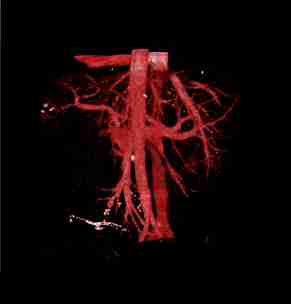 |
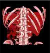
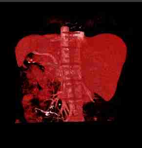
This example concern the volume rendering of a medical image. The windowing range is crucial to improve perception of a region (organ). In this example, the aim is to display venous system from the left figure, knowing that the bones are segmented. Knowing that the hepatic vessels is not included in bones (a priori topologic information), we can remove the bones from the image (middle figure). Doing the hypothesis that the hepatic vessels is more bright than the liver we can update the windowing in order to show only the hepatic vessels (right figure).
Steps |
 |
The steps can be resume by the following items :
- Formalization
- - representation of conceptual information
- - segmentation process modeling
- Inference engine (deduction)
- - region Of Interest
- - number of classes et ordering
- Image
- - masking
- - region clustering
- - optimal windowing
- Evaluation
- - synthetic images
- - clustering algorithm
- - quantification of benefits
- Application
- - medical images
- - cluster identification
- - windowing and volume rendering
Results |
 |
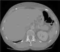
Following the quantitative method evaluation proposed on synthetic images, it been applied on medical images. These data are composed of image and mask (DICOM). The example below shows an image DICOM from X ray scanner, liver and kidney masks.
 |
 |
 |
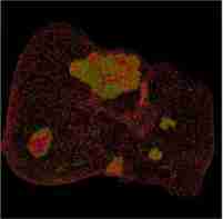
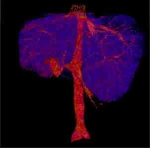
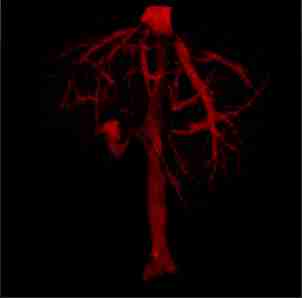
The more significant results from the method are the volume rendering below, it shows liver tumors (left figure) and hepatic vessels in liver (middle figure) and hepatic vessels (right figure) :
 |
 |
 |
Conclusion |
 |
A priori knowledge use confirm, according to the quantitative analysis of the benefits, the interests of the proposed method for both synthetic and medical image. There is an interest obviously to use a priori knowledge for image interpretation. One important point of this success seems to be masking in order to focus a region with a minimum of polluting data. Computing time is then saved. Segmentation performance is improved by a priori number of classes, this allows to identify precisely each class in order to apply an optimal windowing to a structure or organ (visualization improved).
